
New High-Tech Equipment Boosts Cutting-Edge Research at the University of Bayreuth
Good news for research at the University of Bayreuth: A state-of-the-art X-ray computed microtomography (µCT) scanner has been acquired under the leadership of Prof. Dr. Diamantopoulos (Soil Physics) and Prof. Dr. Laforsch (Animal Ecology) as part of a DFG major equipment grant. The new instrument offers fascinating insights into the hidden world of soils and the inner workings of organisms and materials.
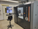
An X-ray computed microtomograph works similarly to a medical CT scanner, but with significantly higher detail resolution. It operates completely without interference and with impressive three-dimensional resolution. Even the finest structures in the micrometer range become visible—from tiny pores in soil to delicate tissue structures in organisms. This allows researchers not only to “look inside,” but also, for the first time, to analyze complex processes within samples under realistic conditions.
“This represents a major breakthrough, especially in soil research,” says Dr. Frederic Leuther of the Chair of Soil Physics. Soils are opaque, and until now, many studies required the destruction of the existing soil structure. With µCT, intact soil samples can now be imaged in full three dimensions. This allows key processes such as water uptake, water movement, and nutrient transport to be studied in their natural environment while preserving the soil structure. The device can also be used for research within the University of Bayreuth’s SFB 1357 Microplastics to address even more realistic research approaches. In combination with existing measurement technology, this also opens up new possibilities for realistic 3D simulations of transport processes in the soil—an important step toward better understanding sustainable land use, the management of water resources, and the transport of microplastics in the soil.
The device also opens up entirely new perspectives in biological research. From changes in organisms under a wide variety of stress conditions to the effects of climate change on animal species: µCT allows for the non-destructive, rapid, and high-resolution 3D visualization of internal and external body structures. This is particularly exciting, for example, for studying larval stages or changes in protective structures such as shells and exoskeletons. Properties such as porosity or material density can also be measured precisely in this way—which is crucial for better understanding how living organisms adapt to changing environmental conditions. At the same time, the new µCT also provides groundbreaking insights into the internal structures of new materials being developed at the University of Bayreuth, which can be improved based on the µCT data.
The significance of this new instrument is also evident in the broad support it has received on the Bayreuth campus: A total of thirteen chairs, the Collaborative Research Centers “Microplastics” (SFB 1357) and “MultiTrans” (SFB 1585), as well as the Bayreuth Center for Ecology and Environmental Research (BayCEER), have endorsed the proposal for this major piece of equipment. The instrument was thus funded with resources from the University of Bayreuth, the SFB Microplastics, and the German Research Foundation (DFG).
The µCT thus establishes a central research infrastructure that bridges disciplines—from environmental and life sciences to materials research and engineering. At the same time, the new technology enhances the University of Bayreuth’s international visibility and opens up numerous new opportunities for collaboration worldwide.

The procurement of the µCT system and the implementation of the project were carried out in collaboration with PHI GmbH, the German sales and service partner of RX Solutions. The collaboration included technical consulting, project coordination, and installation of the system, as well as future technical support. Installing the research device—which weighs about five metric tons and is roughly the size of a wardrobe—posed a particular logistical challenge: The system was hoisted into the temporarily opened building by crane. The spectacular operation generated a great deal of interest on campus, with numerous onlookers watching in amazement as the new large-scale device made its way into the laboratories.
One particular aspect further underscores the project’s international ties: The device’s manufacturer, RX Solutions, is based in Chavanod near Annecy (France)—the very region from which Bayreuth’s sister city hails. Fittingly, Annecy and Bayreuth are celebrating the 60th anniversary of their sister city partnership this summer.

Jennifer Opel
University of Bayreuth
Phone: +49 (0)921 - 55 5893 57
Mail: jennifer.opel@uni-bayreuth.de